One of the many incredible benefits of vagus nerve stimulation (VNS) that some people experience is a boost in their mood. In fact, people with treatment-resistant depression, anxiety, and PTSD have been using medical VNS for years with excellent results. To better understand why these effects are possible, it's helpful to have a comprehensive understanding of the vagus nerve, and that's what we're going to provide you today as we explore the anatomy of the vagus nerve in detail.
The Vagus Nerve
Before we get into the gross anatomy of the vagus nerve, let's begin with an overview of what it is, what it does, and how it does what it does.
The vagus nerve is the longest in the body, originating from the brainstem and extending into the abdomen. It has ten terminal branches, and once you read about them, the function(s) of the vagus nerve becomes a lot more apparent:
- Meningeal
- Auricular
- Pharyngeal
- Carotid body
- Superior laryngeal
- Recurrent laryngeal
- Cardiac
- Pulmonary
- Esophageal
- Gastrointestinal
The vagus nerve has multiple functions, including regulating heart rate, digestion, breathing, salivation, sweating, swallowing, and speaking. It also plays a vital role in keeping our moods stable and helping us to stay relaxed and calm. Based on all of the branches of the vagus nerve, you can start to see how things like heart rate, speech, and digestion can be connected to it.
Gross Vagus Nerve Anatomy
Now let's get into the finer details of vagus nerve anatomy.
Specifically, we'll tell you about the following:
- Cranial nerves
- Vagus nerve branches in the jugular fossa
- Branches in the neck
- Branches in the thorax
- Branches in the abdomen
Cranial nerves
The vagus nerve is the longest of the twelve cranial nerves. Its main function is to send information from the brain to the organs in order to regulate them.
The other eleven cranial nerves are the olfactory nerve (I), optic nerve (II), oculomotor nerve (III), trochlear nerve (IV), trigeminal nerve (V), abducens nerve (VI), facial nerve (VII), vestibulocochlear nerve (VIII), glossopharyngeal nerve (IX) vagus nerve, accessory or spinal accessory nerves, and hypoglossal nerve.
Here's a quick recap of each of these cranial nerves:
- Olfactory nerve (I): Responsible for the sense of smell.
- Optic nerve (II): The sensory nerve that carries visual information from the eye to the brain.
- Oculomotor nerve (III): Motor nerve responsible for controlling eye movements, accommodation, and pupillary constriction.
- Trochlear nerve (IV): Motor nerve responsible for controlling the superior oblique muscle of the eye.
- Trigeminal nerve (V): Both motor and sensory, this is a major pain pathway in humans.
- Abducens nerve (VI): Responsible for lateral movement of the eyeball.
- Facial Nerve (VII): Used to control facial expression and taste sensation on the tongue.
- Vestibulocochlear nerve (VIII): Responsible for balance and hearing sensation.
- Glossopharyngeal nerve (IX): Motor and sensory nerve responsible for controlling the pharynx, larynx, and soft palate, as well as transmitting taste sensations to the brain.
- Vagus nerve (X): Responsible for regulating autonomic functions and transmitting sensory information from the vagus nerve branches.
- Accessory or spinal accessory nerve (XI): Motor nerve responsible for controlling neck muscles and shoulder movement.
- Hypoglossal nerve (XII): Motor nerve responsible for controlling the tongue muscles.
Notably, while many of these cranial nerves serve specific functions, the vagus nerve has many functions. As we explore the anatomy of the vagus nerve, it will become clear just how extensive this nerve and its functions genuinely are.
Vagus nerve branches in the jugular fossa
The vagus nerve begins at the jugular fossa, a shallow depression located at the base of the skull. Here, it has two main terminal branches: meningeal and auricular.
The meningeal branch is a branch of the vagus nerve responsible for supplying the dura mater with sensory information. In contrast, the auricular branch provides sensory innervation to various structures in the ear.
Vagus nerve branches in the neck
The vagus nerve also has four branches that pass through the neck: the pharyngeal branches, superior laryngeal nerves, recurrent laryngeal nerves, and the superior cardiac nerves.
- Superior cardiac nerve: This branch passes through the vagus nerve to supply parasympathetic innervation to the heart. This 2021 paper explains: "While the vagus nerve is within the carotid sheath, it gives off the superior cardiac nerve and is associated with parasympathetic fibers and travels to the heart."
- Pharyngeal branch: This supplies motor innervation to the throat muscles, including the pharynx. It also provides autonomic innervation to some of these muscles.
- Superior laryngeal branch: This vagus nerve branch supplies motor innervation to the cricothyroid muscle, which controls the pitch of your voice and autonomic innervation to other structures in the larynx.
- Recurrent laryngeal branch: This branch supplies motor innervation to all muscles in the larynx except for the cricothyroid and autonomic innervation to the laryngeal structures.
Vagus nerve branches in the thorax
There are also four vagal branches in the thorax: the inferior cardiac nerve, anterior bronchial, posterior bronchial, and the esophageal branches.
- Inferior cardiac nerve: This vagal branch supplies parasympathetic innervation to the lower portion of the heart.
- Anterior and posterior bronchial branches: These vagal nerves supply autonomic innervation to the bronchi to regulate their activity.
- Esophageal branches: These vagus nerve branches provide both motor and autonomic innervation to the esophagus so it can contract and expand appropriately during swallowing.
The left and right vagus nerve also begin to take different paths at this point.
Vagus nerve branches in the abdomen
Finally, the vagus nerve also has all-important branches in the abdomen: the gastric, celiac, and hepatic branches.
The celiac branch supplies vagus nerve fibers to the celiac ganglion, sending them to various abdominal organs, including the pancreas and spleen. For example, vagus nerve fibers supply motor and autonomic innervation to the stomach as well as gastrointestinal smooth muscle. This relates to the gut-brain axis, which is a critical vagus nerve pathway that is involved in its ability to improve mood and symptoms of mental health disorders.
Read more about this gut-brain and vagus nerve connection here.
The hepatic branch provides parasympathetic innervation to the liver, gallbladder, and biliary tract to perform their respective functions.
The gastric branches are worth explaining in a bit more detail:
"The branches of the right vagus nerve forms the posterior gastric plexus on the posteroinferior surface of the stomach, while the branches of the left vagus nerve forms the anterior gastric plexus on the antero-superior surface of the stomach. Both of the divisions run between the layers of lesser omentum.
The fibers from the anterior gastric extend as far as the pylorus and the upper part of the duodenum, while posterior vagal trunk, in addition to posterior gastric branches, sends fibers to major abdominal autonomic plexus from which vagal fibers are distributed to the territories of celiac, renal and superior mesenteric arteries."
Microscopic Anatomy of the Vagus Nerve
With the gross anatomy of the vagus nerve out of the way, let's look at the vagus nerve in even greater detail, starting with its microscopic anatomy.
As this 2015 report explains, the vagus nerve carries five different fiber types: general somatic afferent, general visceral afferent, special visceral afferent, general visceral efferent, and special visceral efferent fibers.
- General somatic afferent fibers convey sensory information about the body (including pain and temperature) to the vagal nerve, which is then transmitted to the brain. On the other hand, general visceral afferents carry sensory information from organs and other body structures.
- Special visceral afferent fibers transmit taste and smell signals from the tongue and pressure signals from muscles in the larynx and pharynx. They develop in association with the gastrointestinal tract.
- General vagal efferents control smooth muscle contractions and gland secretions throughout the body. In contrast, special vagal efferents control involuntary movements of upper airway structures such as the vocal cords, larynx, tongue, and pharynx.
Distribution of Vagus Nerve Fibers
The vagus nerve transmits its fibers to many different parts of the body. Again, as the longest of the cranial nerves, the vagus nerve has a wide distribution throughout the body and is involved in many important physiological functions.
Its main targets are the diaphragm, heart, lungs, gastrointestinal tract, and liver. It also supplies vagal fibers to structures of the neck, such as the larynx and pharynx, for speech production and swallowing. The vagus nerve also supplies autonomic innervation to various organs, such as:
- Pancreas
- Gallbladder
- Urinary bladder
- Adrenal gland
- Spleen
- Multiple muscles in the esophagus, stomach, and intestines
Cough Reflex and Respiration
Another interesting vagus nerve function is its role in the cough reflex. Stimulation of vagal afferents, a branch of vagus nerve fibers located within the respiratory tract, triggers a coughing response.
This response is mediated through vagal pathways, which activate various muscles in the larynx and pharynx to create an airflow that can expel foreign particles from the respiratory system. The vagus nerve thus plays an important role in defending our lungs against invasion by bacteria or other hazardous substances.
The vagus nerve also helps regulate respiration by sending signals to nearby muscles responsible for controlling the rate and depth of breathing. This makes it an integral part of the autonomic nervous system's control over cardiovascular and respiratory functions.
Vagus Nerve Clinical Studies
There are numerous ongoing vagus nerve studies, with a particular focus on how vagal stimulation can help treat mental health disorders.
A few clinical trials currently underway include:
- A Study of Non-invasive Vagus Nerve Stimulation for the Prevention of Migraines
- Pivotal Study of VNS During Rehab After Stroke (VNS-REHAB)
- A Study to Evaluate Feasibility of Microburst Vagus Nerve Stimulator (VNS) Therapy
- A Study of Vagal Nerve Stimulation and Glucose Metabolism
There are also FDA-approved uses for VNS, including treating epilepsy and depression. As research continues to uncover new insights into vagus nerve anatomy, there may soon be even more applications for vagal stimulation as a potential therapeutic option. In the meantime, however, vagal stimulation remains a powerful tool for promoting mental health and wellness.
Conclusion
The vagus nerve is a complex network of nerves that plays a crucial role in our physiology. Its anatomy is responsible for both motor and sensory functions, allowing it to play a key part in numerous bodily activities. It also has implications for mental health, with vagal stimulation used as a treatment for various psychological disorders. As research continues to reveal more about vagus nerve anatomy, we can hope to uncover even more ways this important cranial nerve can be harnessed to promote physical and mental health.
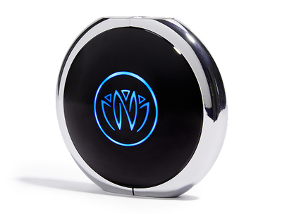
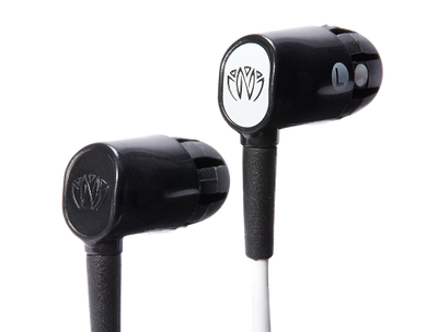
To try VNS for yourself, give the Xen by Neuvana vagus nerve stimulation device a try. It allows you to explore the benefits of vagus nerve stimulation in a non-invasive and comfortable manner.


